

Female infanticide, yes, is a very cruel term wherein the sex of an unborn child is found out and if she is a female child, abortion takes place. Though it is illegal still happens. and census data shows.
The main culprit was the sonography process which identifies the sex of the child and then the parent takes necessary action. Due to those harsh laws has been passed but still, the practice happens. But sonography or ultrasound is not restricted to that only.
From the time it has been used for medical purposes sonography has assisted in helping identify many problems faced by patients like a kidney stone or any other cyst and more. Modern medicine is only benefited from the use of sonography in diagnostic care.

What is Sonography test?
Sonography or Ultrasound was used for a medical purpose from 1956 in Glasgow. The idea of Ultrasound came to two people called Tom Brown who was an engineer and Ian Donald who was an Obstetrician. They created a prototype wherein the instrument created was used to detect the flaws of the industrial ships.
By using high-level ultrasound waves and scanning the internal organs by bouncing the wave on the organ and getting a live image of the scanned area that image is a sonogram. This is similar to how the sonar works and as sound is used for the process it is a safe procedure as no radiation is passed through the body.
How sonography is done?
Preparation before sonography.
Normally sonography or ultrasound is prescribed by the doctor and also mentions the area where sonography or ultrasound has to be done also to do fasting for 8 to 12 hours. Depending upon the area further instruction is given. For example, if the areas are liver, gallbladder, spleen, or pancreas you may be told to eat fat-free food before the fasting period mentioned to you which is normally 12 hours or so.
In the case of other areas, you have to drink a lot of water. Normally there is always this question asked by all of the patients that how much water to drink before going to sonography and It is said you have to drink a minimum of 1 liter or 32 ounces or four glasses or more.
How sonography is done?
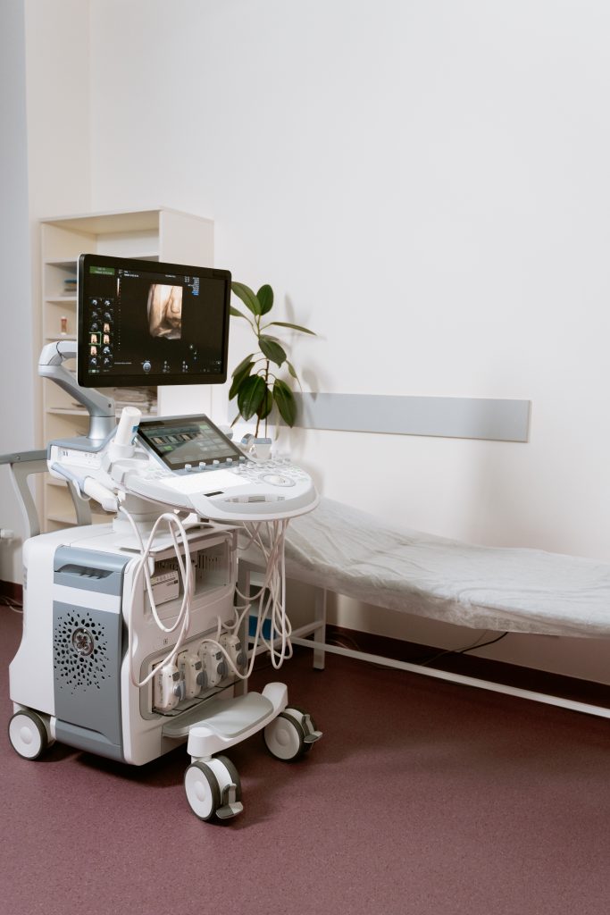
Now, how sonography is done? For performing sonography or ultrasound the device is called a transducer. This transducer is used on the top of the skin to send the ultrasound waves inside the body to the organ which needs to be scanned and listen for the echo.
Before the ultrasound is started a gel is put on the area where the sonography or ultrasound is done. This gel is water-based and propylene glycol and it is a conductive medium and assists in getting the sound waves to travel easily and get a clear picture.
This whole process is captured by the computer and an image is formed based on the echo. This gel is used because ultrasound wave has a hard time traveling through the air and this gel act as a medium to assist the sound wave travel easily.
Many times, the front of the body and back of the body is scanned that is to take the image of the organ from all side to get a clear image of the organ. The same can be printed and seen by the radiologist later on and after doing the sonography or ultrasound report is written and submitted along with the image.

Types of Sonography
Endoscopic ultrasound:
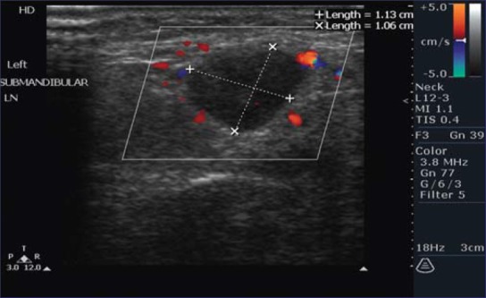
Endoscopic Ultrasound (EUS) is used to assess gastrointestinal or digestive and lung diseases with a minimally invasive procedure. A unique endoscope produces comprehensive pictures of the lining and walls of your digestive system and chest, as well as surrounding organs such as the pancreas and liver, and lymph nodes, using high-frequency sound waves.
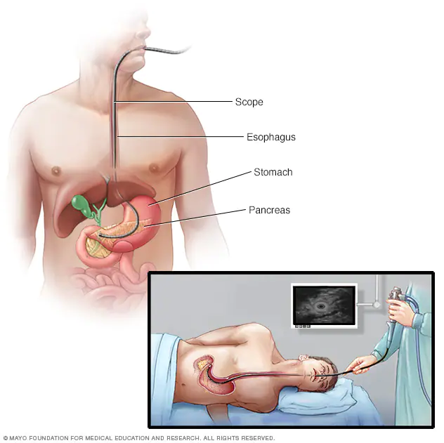
When paired with a fine-needle aspiration technique, EUS allows your doctor to collect (biopsy) fluid and tissue from your abdomen or chest for analysis. EUS combined with fine-needle aspiration may be a less invasive alternative to exploratory surgery.
Doppler ultrasound:
A Doppler ultrasound is a non-invasive diagnostic that uses high-frequency sound waves (ultrasound) to bounce off circulating red blood cells to measure blood flow via your blood arteries. A normal ultrasound produces pictures by using sound waves, but it cannot reveal blood flow.
A Doppler ultrasonography can measure the pace at which blood flows by detecting the rate at which its pitch changes (frequency). A technician trained in ultrasound imaging (sonographer) presses a tiny hand-held instrument (transducer) roughly the size of a soap bar against your skin across the region of your body being scanned, moving from one area to another as needed during a Doppler ultrasound.
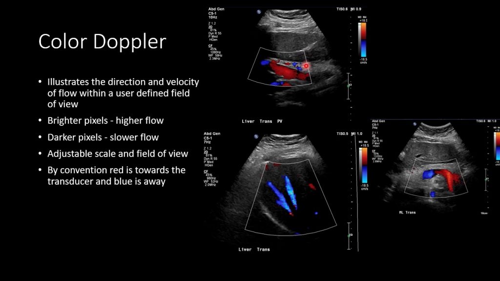
Color Doppler:
A color doppler ultrasound test is a type of ultrasound test where high-frequency sound waves are transmitted into your body and the way the sound bounces back outlines the images on a display. The color doppler test is used to identify the existence of blood clots in the body, as well as obstructions in the arteries and the presence of Deep Vein Thrombosis. The color is superimposed on the image to show the direction and speed of the flow of blood.
Duplex ultrasound:
Duplex ultrasound is used to check the speed of blood flow and the structure of leg vines by using ultrasound waves. Duplex here means that it uses two modes i.e., Doppler and B-mode. B-mode get the image of the vessel and Doppler checks the direction and velocity of the blow flow of the vessel.
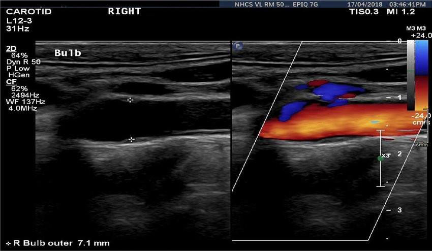
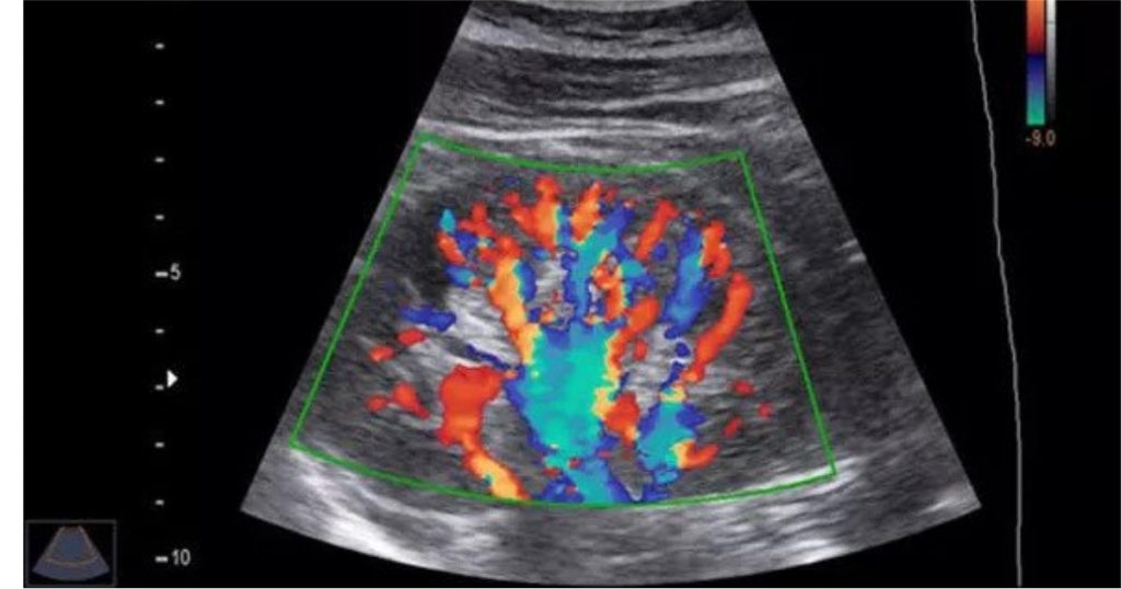
Triplex ultrasound (color-flow imaging):
Triplex ultrasound is also known as color-flow imaging is an upgraded form of Doppler ultrasound. It works the same as Duplex ultrasound but the color is used to show the direction of blood flow. Red color for blood flowing in one direction and blue for another direction.
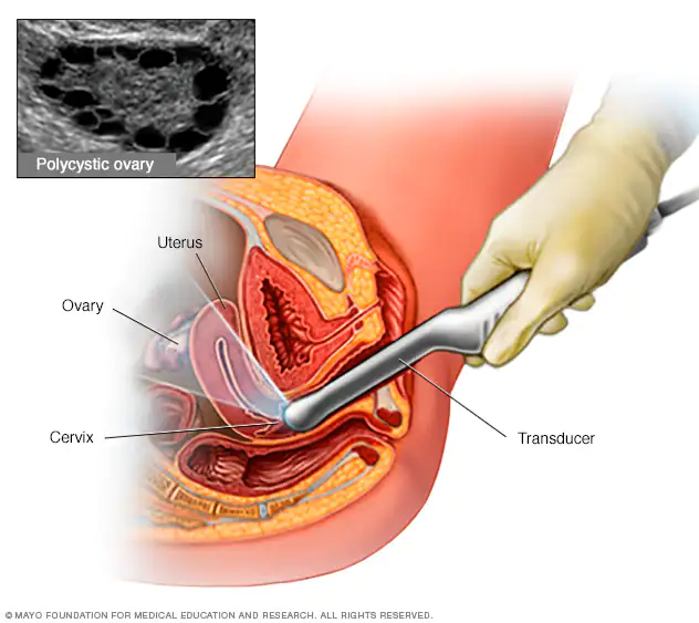
Transvaginal ultrasound:
In Transvaginal Ultrasound to evaluate the endometrium structure, the transducer is inserted in the vagina, and an image is taken to study further to check diseases in the endometrium like adenomyosis, endometriosis, endometrial hyperplasia, and more.
Why sonography is done?
In modern times when sonography is said there are two views, one when a doctor is actually prescribed the same to understand the issue and then to go ahead with diagnosis, and the other is in regards to pregnancy.
Let us understand the first view where the doctor prescribes the sonography to understand the illness or a situation the patient is in. Mainly sonography is done to see the abdomen without actually operating the same. One can see a kidney for kidney stone which can be traced to the urinary tract and see if any stone is blocking it and the dimension of the stone and number of stones.
Sonography is also done for gallstone or gallbladder disease. A gallstone can be of golf ball size or small as a grain of sand and they are made of crystallized cholesterol or pigment or a combination of both. They are usually operated to remove it. Gallbladder disease can be pain on the upper right side of the abdomen which is where the gallbladder is situated.
Another reason is Liver disease. To check how the liver is functioning from Hepatitis to cancer can make the liver function imperfectly and cause problems.
Appendicitis is also another area where sonography is used. The inflammation of the appendix causes pain in the abdomen and it can severely pain and can’t be said reason of pain, for that reason most of the time sonography is done to understand why the pain is caused and where to move forward with treatment.
Overy and Uterus is another major organ where sonography is done for women which comes under pelvic sonography. To find ovarian cyst and uterine fibroids. On the other hand, in males, testicular lumps or testicular cancer are where sonography is used to find the same.
Now coming to the most common use of sonography is to monitor the development of the fetus and uterus during pregnancy time. The most common question is how many sonography done in pregnancy? To answer this mainly 2 times if everything is normal. The first is ideally done in the first trimester to know the due date and the second one at 18-22 weeks to confirm normal anatomy. If abnormalities are found then only repeated sonography or ultrasound is done.
As pregnancy is a delicate matter people often want to know which sonography is required in pregnancy? A transvaginal ultrasound is done to evaluate the condition of the baby and it is endovaginal sonography or ultrasound.
How to read sonography image.
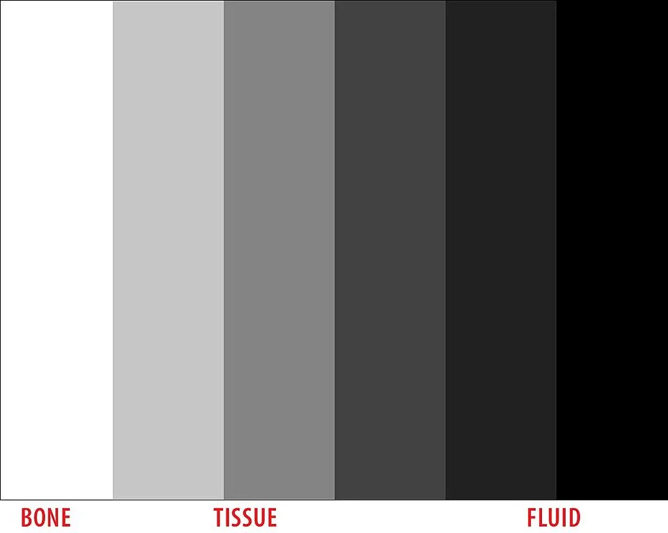
To understand the image fully one needs to study and become a radiologist or a technician but the basic understanding of the sonography image is that fluid is black and tissues are grey and the white is bones. Many times, on the image, is marked the size of deformative structure and distance between them if multiple deformities are found. The same will be mentioned in the written report. If studying them together one can understand the whole situation much better.
Is there any side effect of sonography?
As sonography or ultrasound use sound and is done externally, it is one of the safest procedures as there is no risk of radiation like other procedure. But people do ask is sonography painful? To answer this, it can be a little painful, discomfort, and minor risk of internal bleeding when internal sonography or ultrasound is done.
Can sonography be done at home?
Sometimes pregnant women don’t want to go to the clinic for sonography and ask can sonography be done at home? To answer that no, generally speaking, diagnostic centres are more hygienic, fit for the said activity, and have a comfortable setting for performing the sonography. Moreover, the sonography machine is not mobile in a way that it is difficult to transport from one place to another place. It can be moved within the diagnostic center but outside the diagnostic centre, it will be troublesome.
Conclusion
To summarize, Hope with this article of what is sonography some light is shed on the said topic. Sonography or ultrasound is used to evaluate and diagnose and treat a wide variety of medical conditions from abdominal areas, pelvic region to eyes and blood vessels. It is also one of the most widely used for checking the development of the fetus during pregnancy. It is one of the painless procedures as well as one of the quickest as it takes around 15 to 30 minutes to get the whole procedure over. Sonography is one of the many services provided by modern diagnostic centers.
What is isoechoic?
Tissue or structures that emit an echo of the same strength as the surrounding structures or tissues, making isolation difficult.
Why is fluid black in ultrasound?
Because fluids, such as water or urine, easily transmit sounds, they seem black or deeper in colour.
What is better sonography or MRI
Even though both are imaging tools but they use different core technology. Sonography can not show actual structures but only soft tissues whereas MRI not only shows soft tissues but bones, muscles and joints too. MRI is more modern and provides quality images.
What is the difference between sonography and CT scan?
Sonography uses high-frequency sound waves to map the area of the scan, whereas CT scans use x-ray to scan from a different angle of the designated area of scan. The major difference is with a CT scan there is an exposure to radiation in the patient.

1 thought on “What is Sonography Test – The result of Ultrasound?”
Pingback: What is a diagnostic center : The Essential Guide